Strength Is Coming Back — But Is the Muscle?
What a New Study Reveals About Recovery After ACL Reconstruction
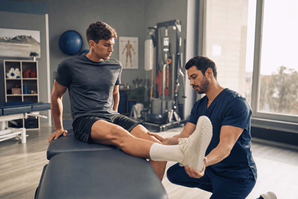
After an ACL reconstruction, most patients and clinicians track progress by what they can feel and do: Is the knee less swollen? Is strength improving? Can I walk without a limp? But a new study asks a more granular question — do strength, muscle size, muscle quality, and self-reported function all move together during early recovery, or do they each tell a different story?
The answer, it turns out, is the latter.
What Was Studied
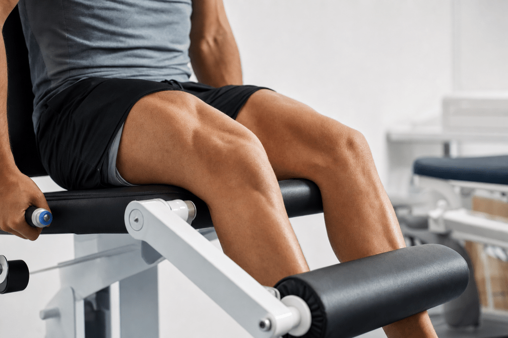
Researchers at the University of Central Florida enrolled 13 participants (4 males, 9 females; mean age approximately 23 years) who had recently undergone ACL reconstruction. The team measured multiple dimensions of recovery — quadriceps strength, muscle cross-sectional area (CSA), muscle quality via ultrasound echo intensity (EI), and self-reported knee function (via the IKDC questionnaire) — at 2, 6, and/or 10 weeks post-surgery, in both the surgical and nonsurgical limbs.
Key Findings
1. The strength gap at 2 weeks is striking.
At the earliest assessment point, the surgical limb produced just 0.19 Nm/kg of isometric quadriceps torque, compared to 3.17 Nm/kg in the nonsurgical limb. That is not a modest deficit — it is near-total functional loss. The good news: surgical-limb strength increased significantly across the 10-week window, while the nonsurgical limb remained stable.
2. Patients felt significantly better over time.
IKDC scores — a patient-reported measure of knee function — started at a mean of just 25.30 out of 100 at 2 weeks. They improved meaningfully and significantly over time, gaining roughly 4 points per week on average.
3. Muscle atrophy continued even as strength improved.
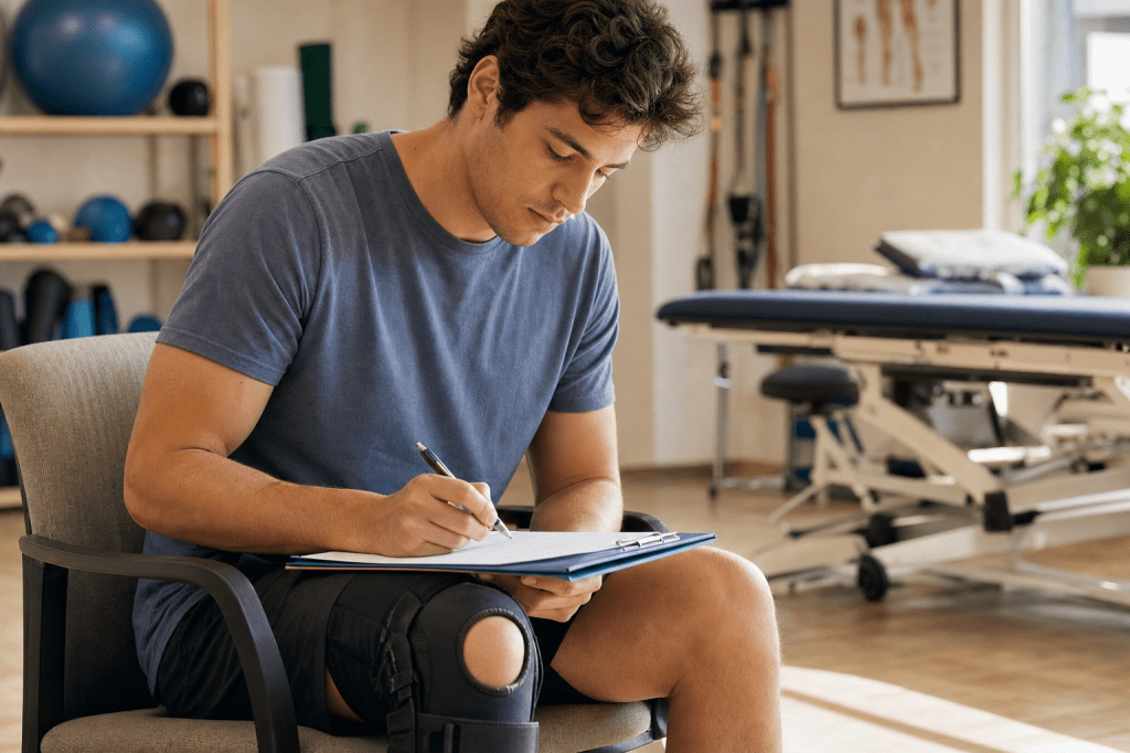
Vastus lateralis CSA decreased significantly in the surgical limb between weeks 2 and 6. In other words, the muscle was still shrinking during a period when strength was already recovering. This dissociation is a notable finding.
4. The rectus femoris showed a rebound — in both legs.
Rectus femoris CSA increased significantly in both limbs between weeks 6 and 10, suggesting a more global muscular response during the later portion of early rehabilitation.
5. Echo intensity did not change — and that itself is a finding.
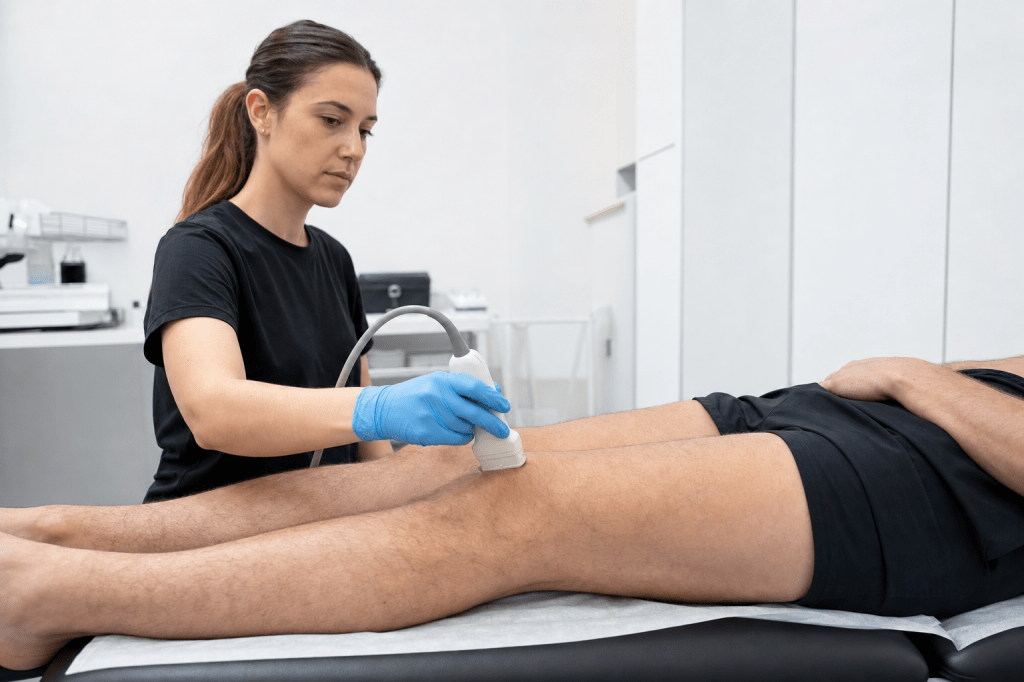
Despite the many changes in strength and size, ultrasound echo intensity — a marker of muscle compositional integrity — did not change significantly over time in either limb, and no between-limb asymmetries were detected at any point. The authors conclude that EI was not a responsive tracking tool during this early window, and suggest it may be better suited for detecting longer-term or persistent compositional changes.
6. None of these measures correlated with each other.
Perhaps the most important finding: changes in strength, size, quality, and self-reported function were statistically independent of one another. Each domain appears to reflect a distinct aspect of recovery — not a single, unified process.
What the Authors Cite From Related Research
The authors note that a 2024 study by Johnston et al. (Medicine & Science in Sports & Exercise) found elevated EI in both the VL and RF following ACLR, with EI and CSA not changing in parallel — supporting the conclusion that EI may lag behind functional improvements. They also reference Garcia et al. (2020, Journal of Athletic Training), who found that quadriceps muscle quality remained impaired at return to activity even after completing rehabilitation, pointing to EI’s potential value over longer timeframes. Additionally, Fasih et al. (2025, International Journal of Sports Medicine) identified substantial EI asymmetries between limbs years after ACLR — which the authors contrast with the early post-surgical period, suggesting EI deficits may not be detectable until recovery extends well beyond 10 weeks.
Limitations to Keep in Mind
The authors are candid about two important constraints. First, the sample was small (N = 13), meaning statistical power was limited and the findings should be treated as exploratory and hypothesis-generating — not definitive. Second, there was no preoperative baseline; assessments began at approximately 2 weeks post-surgery, so it is not possible to fully quantify how much was lost relative to each participant’s true pre-injury status.
The Takeaway
Recovering from ACL reconstruction is not a single process — it is several processes running simultaneously, and they do not necessarily stay in sync. Strength can return while muscle tissue is still shrinking. Patients can feel better before objective measures fully recover. And a tool like echo intensity, while potentially valuable for detecting chronic deficits, may not be sensitive enough to track what is happening in the first 10 weeks.
For clinicians and researchers, that complexity is worth taking seriously.
Disclosure: This blog post is based exclusively on the following peer-reviewed article and reflects only the findings and conclusions of that study. It does not represent a broader literature consensus and should not be used as the basis for clinical decision-making.
Stock MS, Fowler HN, Ditmyer AL, Nyberg CE, Hahs-Vaughn DL, Richardson RM. A Comparison of Methods for Tracking Muscle Quality During Early-Phase Rehabilitation Following Anterior Cruciate Ligament Reconstruction. Journal of Functional Morphology and Kinesiology. 2026;11(2):200. DOI: 10.3390/jfmk11020200


Leave a Reply